Well a bit of a sad day today. We had a Lulu a 10 year old Labrador come in for xrays as she has had an ongoing cough for the last few months. Initially when she came in for this problem around a month ago – she had all the signs and findings of Bordetella bronchiseptica infection (otherwise known as Kennel cough!) and she was treated for this. With no improvement after a 2 week course of an appropriate antibiotic, the decision was made to take some chest xrays.
Chest xrays help us differentiate a lung problem from an upper airway problem and from a heart problem! Large breed dogs, particularly older labs, quite commonly get an upper respiratory problem called ‘Laryngeal paralysis’ – where the larynx (in the throat) doesn’t open as wide as it should – often leading to coughing and hoarse breathing. Lung problems that could cause a cough include anything from asthma, infection, lungworm, pneumonia, cancer and many more! Heart problems include the common dilated cardiomyopathy which causes a dilated heart and thin walled heart muscle, heartworm, cancer and again, many more. This is why getting a least 2 – 3 chest films is so important.
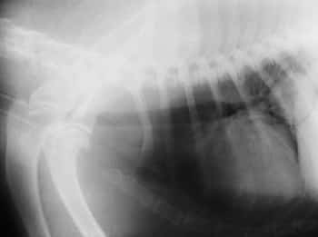
Lulu had 4 different chest xrays taken today under a general anesthetic (to ensure she didn’t move – which would cause a blurred image). The xrays revealed a large abnormal mass in the lung.
Please have a look at the xray from today below (the quality isn’t as good as I took a photo with my digital camera)! Can you spot the mass? The heart here is definitely bigger than usual. We measure the heart a special way by adding the width and height compared to the vertebrae above the chest (the thoracic vertebrae). This gives us a special score (the VHS – vertebral heart score) that allows us to assess the heart size of ALL breeds of dogs. Lulu definitely has heart enlargement which could be contributing to her cough – and so she will be sent for an echo (ultrasound of the heart) very soon.
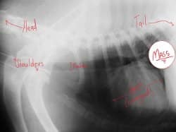
Did you find the mass? Do you know what end of the picture the head would be? Where is the trachea (windpipe)? Have a look at the image below for more help! (Sorry! I had to use ‘paint’!)
If you switch between the two images you can very easily spot the abnormal mass up behind the heart. This area is usually approximately the same black color that you can see to the left of the heart. The question is – what is this mass? Is it a tumor? It is a localized infeciton (aka – an lung abscess)? The mass was visible in the same spot in all four xrays confirming it’s place within the chest in a certain part of the lungs – the right caudal lung lobe. Given lulu’s age and how the mass looks on the xray – it is very much likely that this a tumor. Even if this is the case – there are even more questions to ask! Is this a primary lung tumor (did it start in the lungs?) or is it a secondary lung tumor (is there a tumor somewhere in the body that has metastasized (spread) to other parts of the body, 
An interesting case – but a sad one in that, this is most likely a lung tumor!
I hope you have learnt something from this blog post – even if it’s just seeing what a dog chest looks like on xray!


